Introduction
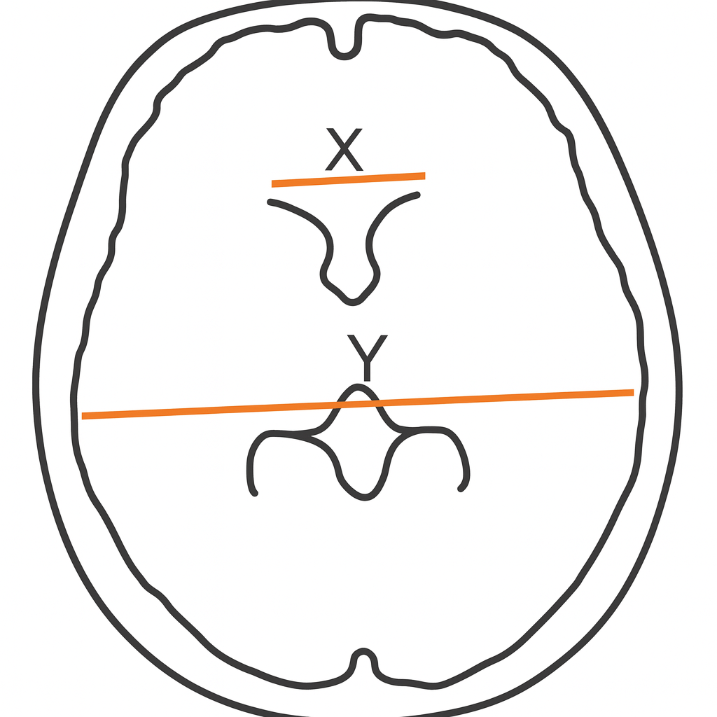
Since its introduction by Evans in 1942, the Evans' Index (EI) - the ratio of the maximal width of the frontal horns to the maximum internal diameter of the skull - has remained one of the simplest radiological tools for assessing ventricular size. Traditionally, a value of >0.30 has been regarded as the threshold for ventriculomegaly and is widely applied in the workup of hydrocephalus, idiopathic normal pressure hydrocephalus (iNPH), and other dementias. However, mounting evidence demonstrates that this single cut-off is inadequate in the elderly, where ventricular enlargement may reflect normal aging rather than pathology.
Limitations of the Classical Cut-Off
Several studies have shown that a significant proportion of cognitively normal elderly individuals exceed the traditional 0.30 threshold without clinical disease. For example, in Brix et al.'s multicenter MRI study of 534 participants, nearly 30% of healthy elderly controls had EI >0.30, underscoring the risk of overdiagnosis when applying static cut-offs in aging populations.
Refined Age- and Sex-Specific Thresholds
The landmark work of Brix et al. (2017, European Journal of Radiology) proposed new age- and sex-stratified cut-offs based on the 97.5th percentile of healthy elderly participants aged 65-84 years:
Age group (years) | Men | Women |
|---|---|---|
65-69 | 0.34 | 0.32 |
70-74 | 0.36 | 0.33 |
75-79 | 0.37 | 0.34 |
80-84 | 0.37 | 0.36 |
These thresholds provided greater specificity (91-98%) for distinguishing iNPH and Alzheimer's disease (AD) from controls compared to the conventional 0.30, though sensitivity remained variable.
Population-Based Normative Studies
Subsequent work has explored normative ranges in different populations:
- Nigeria (Ominde et al., 2025, Nigerian Postgraduate Medical Journal): CT study of normal adults reported an EI range of 0.20-0.34 with means around 0.25. The authors proposed 0.25-0.30 as "borderline" and >0.30 as abnormal.
- Indonesia (Zamzami et al., 2023, Journal of Social Medicine): Mean EI of ~0.26 (±0.03) was observed, with no association between EI and education level. Some regional studies (e.g., Dhok et al. in India) proposed cut-offs up to 0.34 in individuals >70 years.
- Saudi Arabia (Alomar et al., 2024, Neurosciences Journal): Local normative data largely supported Brix et al.'s observations, emphasizing the need for population-specific standards.
Role of Education and Cognitive Reserve
Despite the well-documented role of education in cognitive resilience, no peer-reviewed studies to date have established Evans' Index thresholds stratified by education level. The Indonesian CT study specifically reported no relationship between education and EI values, suggesting ventricular size is determined primarily by age, sex, and biological factors rather than cognitive reserve.
Practical Guidance for Clinicians
- Do not apply a rigid EI >0.30 threshold in elderly patients. Consider age- and sex-specific ranges, particularly those proposed by Brix et al. (2017).
- Interpret EI in context with other imaging markers (callosal angle, disproportionately enlarged subarachnoid space hydrocephalus [DESH] features, volumetric analysis) and the clinical picture.
- Population variability matters. Clinicians should be aware that normative values differ across regions and ethnic groups.
- Education level is not a factor. Current data do not support modifying EI thresholds based on cognitive reserve or years of schooling.
Conclusion
The Evans' Index remains a useful, rapid tool for the radiological assessment of ventricular size. However, reliance on the traditional >0.30 cut-off risks overdiagnosis in older adults. The most robust contemporary data (Brix et al., 2017) provide refined, age- and sex-stratified thresholds that improve specificity in elderly cohorts. Until further large-scale, multiethnic studies refine these standards, clinicians should interpret EI flexibly and always in conjunction with the clinical syndrome and complementary imaging markers.
References
- Brix MK, et al. The Evans' Index revisited: New cut-off levels for use in radiological assessment of ventricular enlargement in the elderly. Eur J Radiol. 2017;95:42-48. doi:10.1016/j.ejrad.2017.07.013.
- Zhou Z, et al. Application of Evans Index in Normal Pressure Hydrocephalus: A Mini Review. Front Aging Neurosci. 2022;14:783092.
- Alomar S, et al. Normative parameters of the Evans Index using Computer Tomography. Neurosciences (Riyadh). 2024;29(2):122-129.
- Ominde BS, et al. Evaluation of the Evans Index on Normal Brain Computed Tomography Scans. Niger Postgrad Med J. 2025;32(1):28-34.
- Zamzami A, et al. Relationship between Evans Index and Demographic Factors in Normal Adults. J Soc Med. 2023;5(2):69-75.
- Radiopaedia.org. Evans' Index. Accessed September 2025.